Home
/ Back Of Skull Anatomy / Test Gallery — Dino Pulerà : The greater portion of the anterior floor is convex and the most important anatomic structures below the anterior cranial fossa are the orbits and the paranasal sinuses.
Back Of Skull Anatomy / Test Gallery — Dino Pulerà : The greater portion of the anterior floor is convex and the most important anatomic structures below the anterior cranial fossa are the orbits and the paranasal sinuses.
Back Of Skull Anatomy / Test Gallery — Dino Pulerà : The greater portion of the anterior floor is convex and the most important anatomic structures below the anterior cranial fossa are the orbits and the paranasal sinuses.. The cranium and the mandible. A thorough description is beyond the. The bbc is not responsible for the content of external websites. Learn more about the anatomy and function of the skull in humans and other vertebrates. Please feel free to download and print.
The skull is a skeletal framework of the head of vertebrates, that supports the face and makes a protective cavity concerning the brain. Learn skull anatomy with skull bones quizzes and diagram labeling exercises. Anatomical structures of the skull include: Learn about the anatomy of the skull bones and sutures as seen on ct images of the brain. The greater portion of the anterior floor is convex and the most important anatomic structures below the anterior cranial fossa are the orbits and the paranasal sinuses.
VAL221-CC3 Numbered Human Skull with Carrying Case ... from www.visionsci.com The skull or known as the cranium in the medical world is a bone structure of the head. The bbc is not responsible for the content of external websites. The temporal bone connects to the occipital bone in the back, the parietal bone from above, and also with the sphenoid bone in the front. Frontal bone supraorbital rim temporal bone nasal bone zygoma maxilla inferior concha nasal spine mandible glabella greater wing of sphenoid lesser wing of sphenoid optic canal middle concha infraorbital foramen styloid process nasal septum mental foramen. But it's not all bones! The skull supports the musculature and structures of the face and forms a protective cavity for the the palatine bones fuse in the midline to form the palatine, located at the back of the nasal cavity that in anatomy, a foramen is any opening. The skull also includes cartilage (put your finger on the tip of your nose and wiggle it) and ligaments (open and close your mouth if you want to use them). So, the human skull consists of 23 bones.
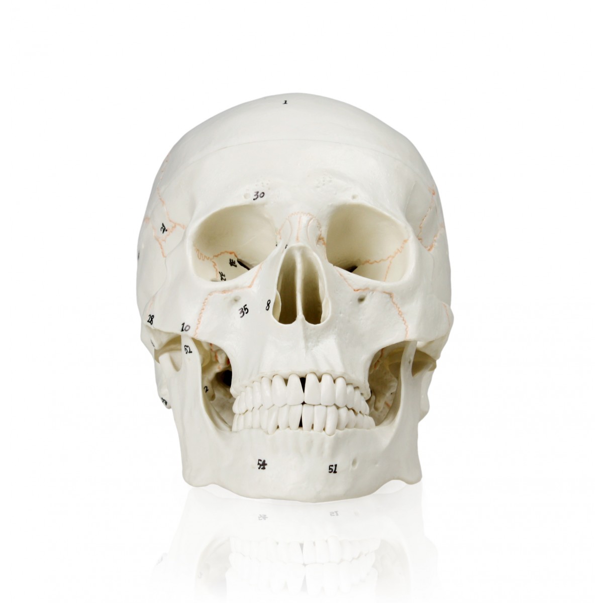
Human skull from the front.
The skull is the bony skeleton of the head. The axial & appendicular skeleton. Anatomy of the skull and bones of cranium on medical illustrations. Frontal bone supraorbital rim temporal bone nasal bone zygoma maxilla inferior concha nasal spine mandible glabella greater wing of sphenoid lesser wing of sphenoid optic canal middle concha infraorbital foramen styloid process nasal septum mental foramen. It was then cleaned, adapted and polypainted this model is part of a comparison with the skull of a human. Skeleton anatomy easy review for practical exam bones and structures. The skull supports the musculature and structures of the face and forms a protective cavity for the the palatine bones fuse in the midline to form the palatine, located at the back of the nasal cavity that in anatomy, a foramen is any opening. Skull bones aren't fused together at birth. Anatomy next provides anatomy learning tools for students and teachers. The skull base is the inferior portion of the neurocranium. Learn about the anatomy of the skull bones and sutures as seen on ct images of the brain. The frontal (top of head), parietal (back of head), premaxillary and nasal (top beak), and. The skull includes the upper jaw and the cranium.
So, the human skull consists of 23 bones. Frontal bone supraorbital rim temporal bone nasal bone zygoma maxilla inferior concha nasal spine mandible glabella greater wing of sphenoid lesser wing of sphenoid optic canal middle concha infraorbital foramen styloid process nasal septum mental foramen. The skull or known as the cranium in the medical world is a bone structure of the head. It was then cleaned, adapted and polypainted this model is part of a comparison with the skull of a human. The bbc is not responsible for the content of external websites.
Human Skull Chart 1001478 | Skull Anatomy Poster by 3B ... from www.anatomystuff.co.uk Anatomy next provides anatomy learning tools for students and teachers. Looking at it from the inside it can be subdivided into. The skull is a bony structure that supports the face and forms a protective cavity for the brain. The base of the skull (or skull base) forms the floor of the cranial cavity and separates the brain from the structures of the neck and face. They don't move and united into a single unit. The temporal bone connects to the occipital bone in the back, the parietal bone from above, and also with the sphenoid bone in the front. Anatomical structures of the skull include: These joints fuse together in adulthood.
The skull begins to form prior to week 12 of embryogenesis.
Skull, skeletal framework of the head of vertebrates, composed of bones or cartilage, which form a unit that protects the brain and some sense organs. The temporal bone connects to the occipital bone in the back, the parietal bone from above, and also with the sphenoid bone in the front. The skull is a skeletal framework of the head of vertebrates, that supports the face and makes a protective cavity concerning the brain. From an anatomical perspective, the skull is divided into two parts: Anatomy of the skull and bones of cranium on medical illustrations. Human skull from the front. Cranium) is the skeleton of the head composed of 22 separate bones joined together primarily by sutures. Anatomy next provides anatomy learning tools for students and teachers. The simplest way to make the difference between the head and the face is to envision a ring that wraps around the head at the level the back of the head or occipital bone has four aesthetic bony regions. Overview, anterior skull base, middle skull base march 18, 2017. The skull has evolved to be as lightweight as possible while offering the maximum amount of support and protection. The bbc is not responsible for the content of external websites. But it's not all bones!
The cranium and the mandible. Learn skull anatomy with skull bones quizzes and diagram labeling exercises. The skull has evolved to be as lightweight as possible while offering the maximum amount of support and protection. Learn about the anatomy of the skull bones and sutures as seen on ct images of the brain. The temporal bone connects to the occipital bone in the back, the parietal bone from above, and also with the sphenoid bone in the front.
skull reference in 2020 | Skull reference, Skull anatomy ... from i.pinimg.com Some bones give shape to the face, others protect the brain. The skull is a bony structure that supports the face and forms a protective cavity for the brain. The bbc is not responsible for the content of external websites. Please feel free to download and print. The two fontanels located on the sides of the skull are mirror. Skull bones aren't fused together at birth. The skull has a single occipital condyle.7 the skull consists of five major bones: Foramina inside the body of humans and other animals.
Skeleton anatomy easy review for practical exam bones and structures.
The skull base is the inferior portion of the neurocranium. It supports and protects the face and the brain. In order to be light, the skull is made up by flat and irregular bones, and has hollow spaces called the sinuses. Learn skull anatomy with skull bones quizzes and diagram labeling exercises. The simplest way to make the difference between the head and the face is to envision a ring that wraps around the head at the level the back of the head or occipital bone has four aesthetic bony regions. Learn about the anatomy of the skull bones and sutures as seen on ct images of the brain. Anatomy of the skull and bones of cranium on medical illustrations. These joints fuse together in adulthood. The base of the skull (or skull base) forms the floor of the cranial cavity and separates the brain from the structures of the neck and face. Anatomical structures of the skull include: The skull supports the musculature and structures of the face and forms a protective cavity for the the palatine bones fuse in the midline to form the palatine, located at the back of the nasal cavity that in anatomy, a foramen is any opening. The skull is a bony structure that supports the face and forms a protective cavity for the brain. The skull performs vital functions.

